Overview
Diagnosing heart failure with preserved ejection fraction (HFpEF) with the HFA-PEFF and H2FPEF scores depends on echocardiographic measurements, which are time-consuming and vary between observers. This study asked whether scores built from fully automated deep learning measurements perform similarly to scores built from manual measurements. It builds on earlier validation of the Us2.ai workflow, including the multicohort study of deep learning in echocardiography.
Study Design
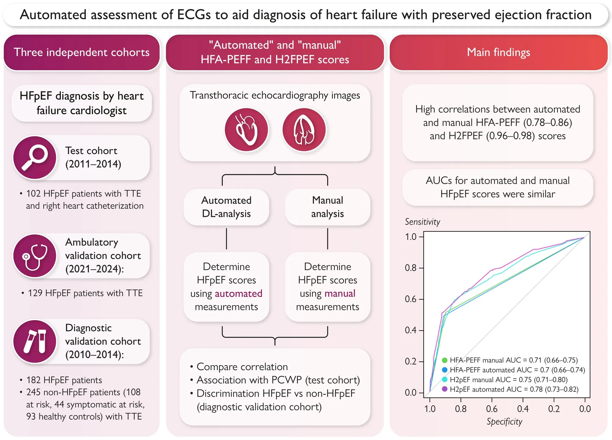
The investigators analyzed echocardiograms from three cohorts. Certified sonographers measured every study manually according to local standards. The same studies were also processed with an automated, validated deep learning workflow (Us2.ai, version 2.2). The algorithms identify the correct 2D or Doppler view, segment structures and provide measurements. HFA-PEFF and H2FPEF scores were then calculated from the manual measurements and from the automated measurements.
- Test cohort: 102 patients with symptomatic HFpEF from a randomized trial of sildenafil (2011 to 2014), all with right heart catheterization and simultaneous echocardiography.
- Ambulatory validation cohort: 129 patients with HFpEF from a dedicated outpatient clinic (2021 to 2024).
- Diagnostic validation cohort: 427 participants from the Alberta HEART study (2010 to 2014), of whom 182 had HFpEF and 245 did not. The non-HFpEF group included 108 patients at risk, 44 symptomatic patients at risk and 93 healthy controls.
Heart failure cardiologists established all HFpEF diagnoses according to European Society of Cardiology guidelines. The investigators compared the scores using correlation, agreement and reclassification between risk categories, correlation with pulmonary capillary wedge pressure (PCWP) in the test cohort, and area under the curve (AUC) for separating HFpEF from non-HFpEF in the diagnostic validation cohort.
Key Results
- Automated and manual scores correlated well across cohorts, with correlation coefficients of 0.78 to 0.86 for HFA-PEFF and 0.96 to 0.98 for H2FPEF. Correlations for individual echocardiographic measurements ranged from 0.43 to 0.93.
- The algorithm returned every echocardiographic component of both scores in 42% of the test cohort, 82% of the ambulatory validation cohort and 49% of the diagnostic validation cohort. The authors attribute missing measurements to poor image quality.
- Both scores correlated similarly with PCWP. For HFA-PEFF, r was 0.23 (automated) and 0.30 (manual), P = 0.25. For H2FPEF, r was 0.33 for both, P = 0.93. The authors describe these correlations as poor for both methods.
- About one in five patients classified as high likelihood by manual HFA-PEFF scores were classified as intermediate likelihood by automated scores (21% of 71 patients in the test cohort and 18.6% of 87 in the ambulatory validation cohort). This was mainly due to lower left atrial volume index estimates by the automated method, and the difference did not consistently vary with atrial fibrillation.
- In the diagnostic validation cohort, AUCs for separating HFpEF from non-HFpEF were similar for automated and manual HFA-PEFF scores (0.70 [95% CI 0.66 to 0.74] vs. 0.71 [95% CI 0.66 to 0.75]). For H2FPEF, the automated AUC was 0.78 (95% CI 0.73 to 0.82) vs. 0.75 (95% CI 0.71 to 0.80) for manual scores, a small but statistically significant difference (P = 0.01).
Why This Matters
HFpEF is hard to diagnose, and the two scoring systems often disagree when their echocardiographic inputs differ. Consistent measurements are therefore central to using the scores. In this study, scores from automated measurements gave similar results to manual scores in three independent cohorts, which supports the feasibility of using automated echo analysis to help with HFpEF diagnosis.
Conclusion
The authors conclude that HFA-PEFF and H2FPEF scores based on automated and manual analysis showed similar diagnostic accuracy, and that automated HFpEF diagnosis using deep learning analysis of echocardiograms is feasible. They note that the findings warrant prospective validation, and that further investigation remains warranted when scores disagree or the likelihood is intermediate. Their stated limitations are that studies were measured according to local standards and not in a core laboratory, that newer and higher-quality images returned more complete automated measurements, that the AUCs may be inflated by incorporation bias and the inclusion of healthy controls, and that left atrial volume differed notably between methods.
Venema, C. S., Venner, M., Achten, A., Ouwerkerk, W., Raafs, A., Mourmans, S., Sikking, M., Rocca, H.-P. B.- La, Heymans, S., van Empel, V. P. M., Hummel, Y., Ezekowitz, J., Hoendermis, E., Voors, A. A., Wykrzykowska, J. J., Knackstedt, C., Tromp, J., & Weerts, J. (2026). Automated assessment of echocardiograms to aid diagnosis of heart failure with preserved ejection fraction. European Heart Journal - Digital Health, 7(8), ztag142. https://doi.org/10.1093/ehjdh/ztag142