Us2.ai Receives CE Mark for Latest AI Echocardiography Capabilities, Unlocking Fully Automated Analysis and Single-View Cardiac Amyloid Detection Across Europe
Us2.ai, a global leader in artificial intelligence (AI) for ultrasound imaging, is proud to announce that its latest version of fully automated echocardiography software has received CE Mark clearance, expanding its clinical deployment across Europe.
This milestone marks a significant advance in Us2.ai’s mission to democratize high-quality echocardiography (ultrasound of the heart). The newly approved version offers comprehensive, guideline-directed echo analysis—entirely automated and instantly reportable—bringing expert-level insights to every echo exam, regardless of operator experience or location.
What’s New in This Release?
Expanded CE-Marked Measurements for Comprehensive Echo Reports
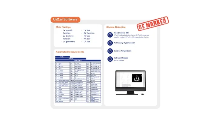
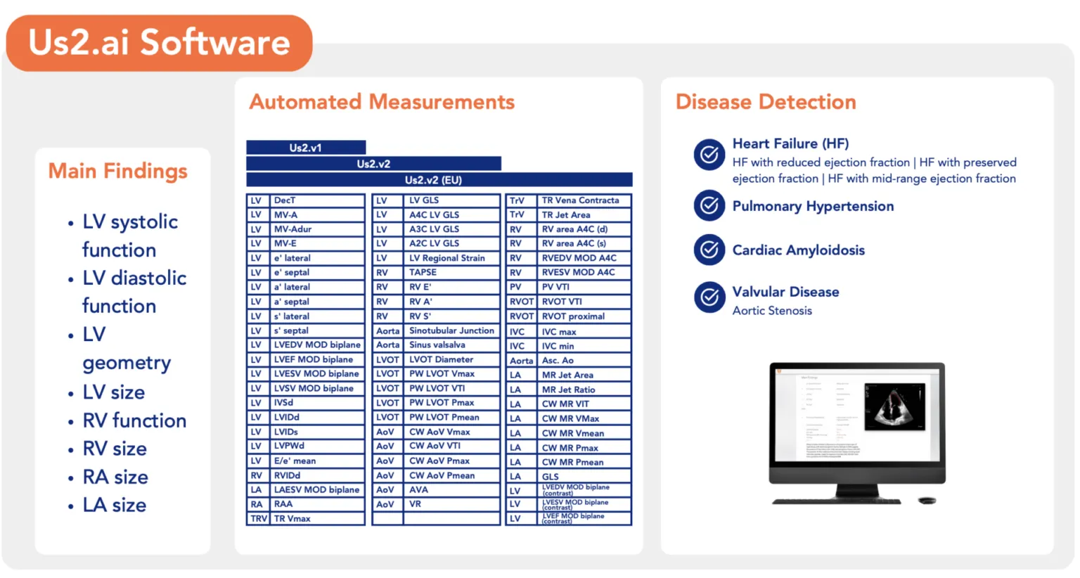
The updated platform now includes 68 CE-marked echo parameters, encompassing cardiac chamber quantification, systolic/diastolic function, valvular assessment, and myocardial strain. All outputs are automatically referenced to international guidelines (ASE, EACVI), enabling fast, accurate, and reproducible diagnostic reports.
Breakthrough: Cardiac Amyloidosis Detection from a Single A4C View
In a first-of-its-kind capability, the software now incorporates AI-based pattern recognition to detect cardiac amyloidosis from a single apical four-chamber (A4C) view [1]—offering clinicians a practical tool for screening a frequently overlooked and deadly condition. This feature, validated in multi-center studies, is now CE-marked and available across Europe.
Clinical-Grade Automation at Scale
Deployed across hospitals and research centers in the Netherlands, Poland, Sweden, France, Germany, and the UK [for use cases, refer to 2–3], Us2.ai’s software is already powering real-world clinical workflows. Clinicians can now access:
- Automated GLS and regional strain, supporting nuanced diagnosis of heart failure and coronary artery disease [4–5]
- Automated Aortic Stenosis severity grading, aligned with EACVI/ASE criteria [6]
- Guideline-compliant HFpEF and Amyloidosis diagnostic parameters [7–9]
- Automated systolic/diastolic function assessment, validated across multi-cohort international datasets [9]
Clinical Perspectives
"This release fully automates the acquisition of all key diagnostic parameters in a guideline-directed echocardiogram. From strain to structure to screening, we are now enabling point-of-care decision support at scale—with accuracy comparable to expert manual review. Us2.ai is truly a co-pilot for echocardiography.” — Yoran Hummel Co-Founder, Us2.ai
“Cardiac amyloidosis remains a frequently under-diagnosed condition with significant prognostic implications. Us2.ai’s AI-based pattern recognition model, applied to a standard apical four-chamber view, provides a validated and scalable solution to support early detection and enhance clinical decision-making within routine echocardiographic practice.“ —Marianna Fontana Professor of Cardiology and Honorary Consultant Cardiologist, National Amyloidosis Centre, University College London
Key Capabilities
- Real-time, fully automated analysis: No manual tracing or measurement required.
- Explainable AI: Transparent visual overlays and editable outputs support confident interpretation.
- Workflow integration: Plug-and-play into existing echo systems, reducing time-to-diagnosis and eliminating reporting bottlenecks.
- Disease screening: Proven applications in community-based screening [10–12].
- Research acceleration: Further proven applications in clinical trials
Why It Matters—for Clinicians, Health Systems, and AI Leaders
- For clinicians: Improve reproducibility, efficiency and accuracy, while reducing fatigue and variability.
- For hospitals: Cut exam and reporting times, optimize resource use, and standardize quality across care levels.
- For innovators and researchers: Provide a scalable, explainable AI framework that enables structured data output for clinical trials, registries, and population health initiatives.
For clinical demos, implementation support, or partnership inquiries, please contact [email protected].
About Us2.ai
Us2.ai is transforming echocardiography through artificial intelligence. Our mission is to make high-quality, guideline-based heart disease diagnosis accessible to everyone—everywhere. The platform is vendor-neutral, fully automated, and backed by peer-reviewed evidence across continents.
References:
[1] Ioannou A, et al. Presented at EuroEcho-Imaging 2024; 11–13 December 2024; Berlin, Germany. https://us2.ai/news/ai-echo-to-diagnose-cardiac-amyloidosis-a-multi-centre-international-development-and-validation-stu
[2] AI Echo in the Netherlands – Us2.ai. (2025). Us2.AI. https://us2.ai/news/ai-echo-in-the-netherlands/
[3] Lafitte, S., Lafitte, L., Jonveaux, M., Pascual, Z., Ternacle, J., Dijos, M., Bonnet, G., Reant, P., & Bernard, A. (2025). Integrating artificial intelligence into an echocardiography department: Feasibility and comparative study of automated versus human measurements in a high-volume clinical setting. Archives of Cardiovascular Diseases. https://doi.org/10.1016/j.acvd.2025.04.051
[4] Myhre, P. L., Hung, C., Frost, M., Jiang, Z., Ouwerkerk, W., Teramoto, K., Svedlund, S., Saraste, A., Hage, C., Tan, R. S., Beussink‐Nelson, L., Fermér, M. L., Gan, L., Hummel, Y. M., Lund, L. H., Shah, S. J., Lam, C. S., & Tromp, J. (2023). External validation of a deep learning algorithm for automated echocardiographic strain measurements. European Heart Journal, 5(1), 60-68. https://doi.org/10.1093/ehjdh/ztad072
[5] Balinisteanu, A., Duchenne, J., Puvrez, A., Wouters, L., Bézy, S., Youssef, A., Minten, L., Bekhuis, Y., van Langenhoven, L., Papangelopoulou, K., Cieplucha, A., Cattapan, I., Tostes, P., Bogaert, J., Vinereanu, D., Thomas, J. D., Badano, L., & Voigt, J.-U. (2025). Vendor Differences in 2D-Speckle Tracking Global Longitudinal Strain: An Update on a Ten-Year Standardization Effort. European Heart Journal – Cardiovascular Imaging. https://doi.org/10.1093/ehjci/jeaf155
[6] Krishna, H., Desai, K., Slostad, B., Bhayani, S., Arnold, J. H., Ouwerkerk, W., Hummel, Y., Lam, C. S. P., Ezekowitz, J., Frost, M., Jiang, Z., Equilbec, C., Twing, A., Pellikka, P. A., Frazin, L., & Kansal, M. (2023). Fully Automated Artificial Intelligence Assessment of Aortic Stenosis by Echocardiography. Journal of the American Society of Echocardiography : official publication of the American Society of Echocardiography, 36(7), 769–777. https://doi.org/10.1016/j.echo.2023.03.008
[7] Cotella, J. I., Randazzo, M., Maurer, M. S., Helmke, S., Scherrer‐Crosbie, M., Soltani, M., Goyal, A., Zaręba, K. M., Cheng, R. K., Kirkpatrick, J. N., Yogeswaran, V., Kitano, T., Takeuchi, M., Fernandes, F., Hotta, V. T., Vieira, M. L. C., Elissamburu, P., Ronderos, R., Prado, A., . . . Lang, R. M. (2024). Limitations of apical sparing pattern in cardiac amyloidosis: a multicentre echocardiographic study. European Heart Journal. Cardiovascular Imaging. https://doi.org/10.1093/ehjci/jeae021
[8] Ioannou, A., Patel, R. K., Razvi, Y., Hanger, M., Martinez‐Naharro, A., Venneri, L., Lim, H. S., Yoran, H., Frost, M. W., Lam, C. S., Gillmore, J. D., & Fontana, M. (2023). Automated analysis of echocardiograms at diagnosis is able to predict prognosis in ATTR cardiomyopathy. European Heart Journal. Cardiovascular Imaging, 24(Supplement_1). https://doi.org/10.1093/ehjci/jead119.387
[9] Tromp, J., Seekings, P. J., Hung, C. L., Iversen, M. B., Frost, M. J., Ouwerkerk, W., Jiang, Z., Eisenhaber, F., Goh, R. S. M., Zhao, H., Huang, W., Ling, L. H., Sim, D., Cozzone, P., Richards, A. M., Lee, H. K., Solomon, S. D., Lam, C. S. P., & Ezekowitz, J. A. (2022). Automated interpretation of systolic and diastolic function on the echocardiogram: a multicohort study. The Lancet. Digital health, 4(1), e46–e54. https://doi.org/10.1016/S2589-7500(21)00235-1
[10] Sankaranarayanan, R., Hartshorne-Evans, N., Mclean, L., Jones, J., Salla, M., Chakrabarti, B., Hadcroft, J., Pritchard, C., Smith, A., & Lam, C. S. P. (2025). Early Detection of Cardiorespiratory Diseases at Everton BEAT-Breathlessness Community Hub: How Football Can Help Save Lives. JACC: Heart Failure. https://doi.org/10.1016/j.jchf.2025.01.008
[11] Hirata, Y., Nomura, Y., Saijo, Y. et al. Reducing echocardiographic examination time through routine use of fully automated software: a comparative study of measurement and report creation time. J Echocardiogr 22, 162–170 (2024). https://doi.org/10.1007/s12574-023-00636-6
[12] Sakamoto, A., Kagiyama, N., Sato, E., Nakamura, Y., Kaneko, T., Miyazaki, S., Minamino, T. (2024). Artificial Intelligence-based Automated ECHOcardiographic Measurements and the Workflow of Sonographer (AI-ECHO): Randomized Crossover Trial. Presented at: AHA 2024. November 16, 2024. Chicago, IL. https://us2.ai/news/ai-echo-rct/